Feature
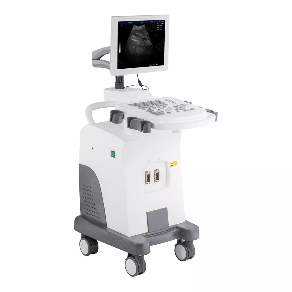
• 15 inch high definition SVGA non-interlaced scanning monitor
• Professional embedded ultrasonic platform,
• High definition full digital image formation technology
• Broadband frequency conversion technology
• To achieve the best combination of high penetration and higher resolution of the organization
• Intellectualized TGC gain control of the whole paragraph 8
• Reliable backlit, silica gel keyboard, photoelectric-tracking control
• Powerful software functions
Rich clinical application
Application to abdomen, uterine attachment, superficial tissue and other clinical tests.
Technical Specifications
|
Scanning mode |
Convex/ linear/ micro-convex |
|
Display |
15-inch LED |
|
Display mode |
B, B+B, 4B, B+M, M. |
|
Detecting depth |
≥244mm |
|
Lateral resolution |
≤2mm (depth≤80mm) |
|
Axial resolution |
≤1mm (depth≤80mm) |
|
Blind Zone |
≤2 mm |
|
Geometry precision |
lateral≤5%, axial≤5% |
|
Gray scale |
256 |
|
Magnification |
×0.8, ×1.0, ×1.2, × 1.5, ×1 .8,
×2.0 |
|
Local zoom |
Yes |
|
Dynamic scope |
0~100dB, visible & adjustable |
|
Electronical focusing |
multi-point or dynamic focusing |
|
Pseudo color |
7 |
|
Measurement |
distance, perimeter, area, heart
rate, gestational week (BPD, CRL, GS, FL, HC, AC, EDD, AFI) etc. |
|
Note |
ID, sex, age, hospital, doctor,
comment, date, time. |
|
Body marks |
30 |
|
Probe interface |
2 |
|
Puncture guide |
The puncture guideline can be
displayed under B mode. |
|
Gain control |
The total gain, 8-segment TGC |
|
Image conversion |
up/ down, left / right, black / white. |
|
Cine loop |
256 frames |
|
Image review |
Images can be reviewed
successively and checked one by one. |
|
Image storage |
40 |
|
Video output |
SVGA (SVGA color monitor), PAL |


